Review sheet anatomy of the respiratory system – Embark on a comprehensive exploration of the respiratory system’s intricate anatomy through this meticulously crafted review sheet. Delving into the structural components and physiological processes that govern respiration, this guide provides a profound understanding of the mechanisms that sustain life.
From the nasal cavity to the alveoli, we will traverse the respiratory system’s anatomical landscape, unraveling the intricate interplay between its various components and their vital role in maintaining homeostasis.
Respiratory System Overview

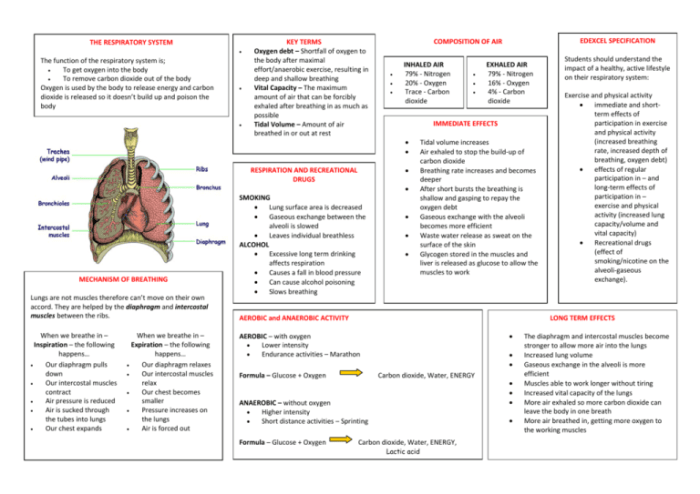
The respiratory system is a complex network of organs and tissues that work together to facilitate the exchange of gases between the body and the external environment. Its primary functions include:
- Providing oxygen to the body’s cells
- Removing carbon dioxide, a waste product of cellular metabolism
Anatomically, the respiratory system is divided into two main sections: the upper respiratory tract and the lower respiratory tract.
The upper respiratory tract consists of the nose, pharynx, and larynx. The nose is responsible for filtering and warming inhaled air, while the pharynx and larynx facilitate the passage of air to and from the lungs.
The lower respiratory tract includes the trachea, bronchi, and lungs. The trachea is a tube-like structure that connects the larynx to the lungs, while the bronchi are smaller tubes that branch off from the trachea and enter the lungs. The lungs are the primary organs of respiration, where gas exchange occurs between the blood and the air.
The respiratory system is closely interconnected with other body systems, including the circulatory system, which transports oxygen and carbon dioxide throughout the body, and the nervous system, which regulates respiratory rate and depth.
Upper Respiratory Tract
Nasal Cavity, Review sheet anatomy of the respiratory system
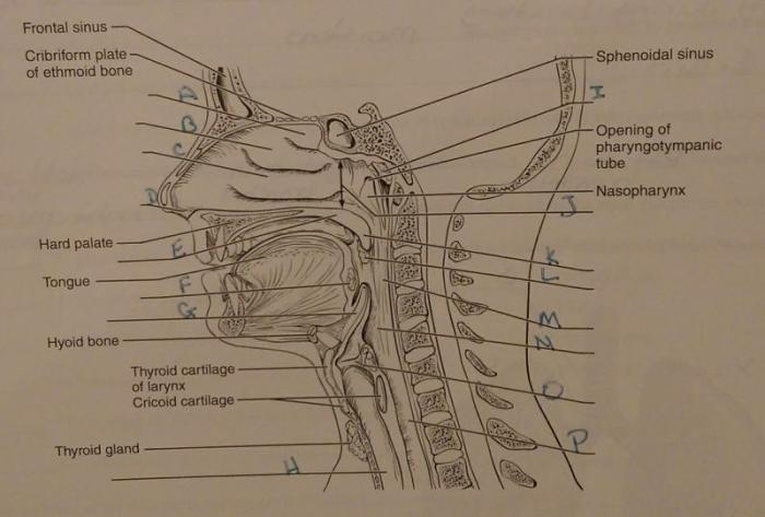
The nasal cavity is a hollow space located behind the nose. It is lined with mucous membranes that help to trap dust, pollen, and other particles from entering the lungs. The nasal cavity also helps to warm and humidify inhaled air.
Pharynx
The pharynx is a muscular tube that connects the nasal cavity to the larynx. It is divided into three sections: the nasopharynx, oropharynx, and laryngopharynx. The nasopharynx is located behind the nasal cavity and is responsible for filtering and warming inhaled air.
The oropharynx is located behind the mouth and is responsible for swallowing. The laryngopharynx is located behind the larynx and is responsible for directing air to and from the lungs.
Larynx
The larynx, also known as the voice box, is a cartilaginous structure located at the top of the trachea. It contains the vocal cords, which vibrate to produce sound.
Lower Respiratory Tract: Review Sheet Anatomy Of The Respiratory System

Trachea
The trachea is a tube-like structure that connects the larynx to the lungs. It is lined with ciliated epithelium, which helps to remove mucus and foreign particles from the lungs.
Bronchi
The bronchi are smaller tubes that branch off from the trachea and enter the lungs. They are lined with ciliated epithelium and mucus-producing glands.
Lungs
The lungs are the primary organs of respiration. They are composed of millions of tiny air sacs called alveoli. The alveoli are lined with capillaries, which allow for the exchange of oxygen and carbon dioxide between the blood and the air.
Alveoli
The alveoli are small, sac-like structures that make up the functional units of the lungs. They are lined with capillaries, which allow for the exchange of oxygen and carbon dioxide between the blood and the air.
Respiratory Cycle

Inhalation
Inhalation is the process of breathing in. It is an active process that requires the contraction of the diaphragm and the intercostal muscles. The diaphragm is a dome-shaped muscle located at the bottom of the chest cavity. When it contracts, it flattens and moves downward, increasing the volume of the chest cavity.
The intercostal muscles are located between the ribs. When they contract, they lift the ribs upward and outward, further increasing the volume of the chest cavity.
As the volume of the chest cavity increases, the pressure inside the lungs decreases. This causes air to flow into the lungs through the nose or mouth, down the trachea, and into the bronchi and alveoli.
Exhalation
Exhalation is the process of breathing out. It is a passive process that occurs when the diaphragm and intercostal muscles relax. As the diaphragm and intercostal muscles relax, the chest cavity decreases in volume, increasing the pressure inside the lungs.
This causes air to flow out of the lungs through the nose or mouth.
Top FAQs
What are the primary functions of the respiratory system?
Gas exchange, providing oxygen to the body and removing carbon dioxide.
Describe the anatomical location of the respiratory system.
The respiratory system extends from the nasal cavity and pharynx to the lungs and alveoli.
What is the role of the diaphragm in respiration?
The diaphragm is the primary muscle of inspiration, contracting to increase lung volume.